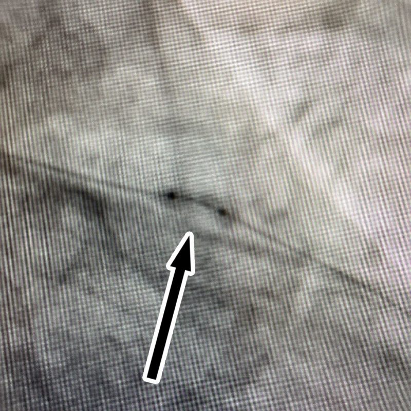
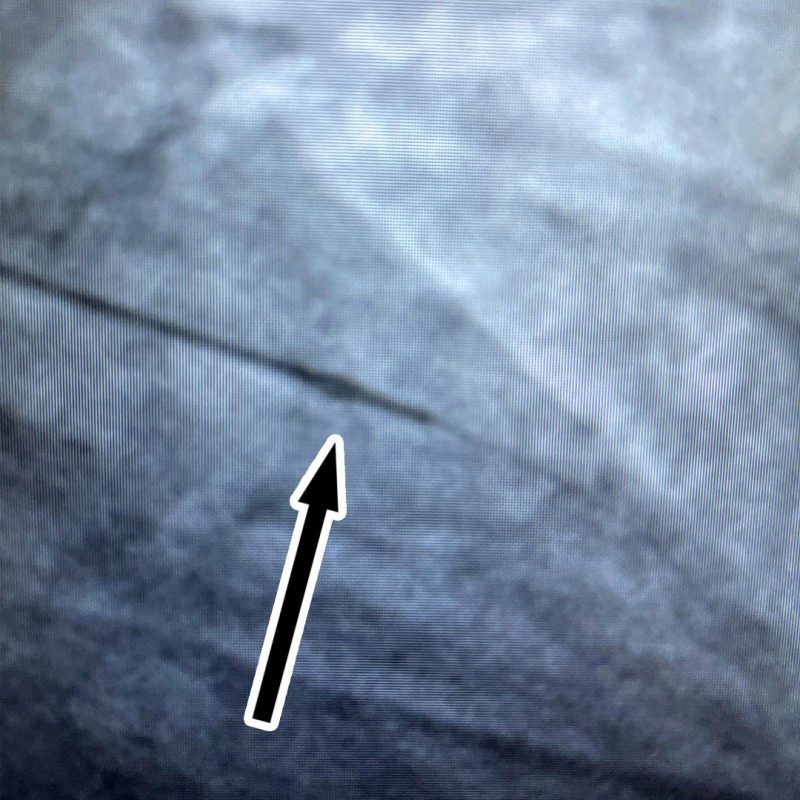
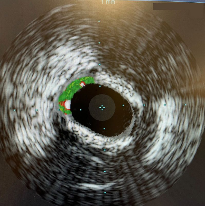
In March of 2021, a 48-year-old male patient had metabolic syndrome and presented with angina pectoris (chest pain). The nuclear stress test in 2019 was normal. However, the coronary calcium score done in 2021 was 1450, which is severely elevated. In March 2021, we took him to the cardiac catheterization laboratory. He had extensive coronary calcification (plaque in his arteries), high-grade lesions noted in the right coronary artery and the two segments, a high-grade lesion in the circumflex trunk, and high-grade stenosis in the proximal left anterior descending artery.

The patient underwent successful stent placement of the right coronary artery and the circumflex artery. Because of the extensive calcification, the lesions were very calcified and noncompliant. This makes the blood vessel very stiff and difficult to expand fully.

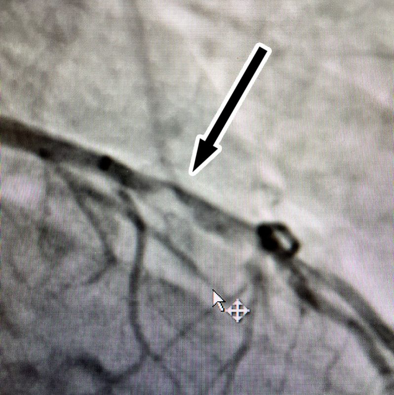
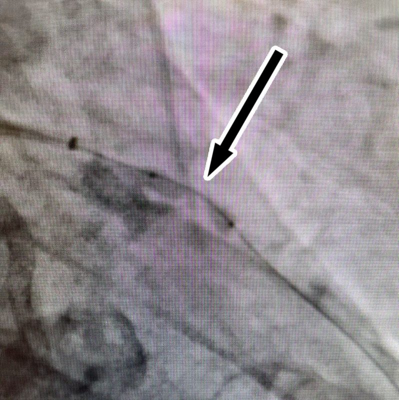
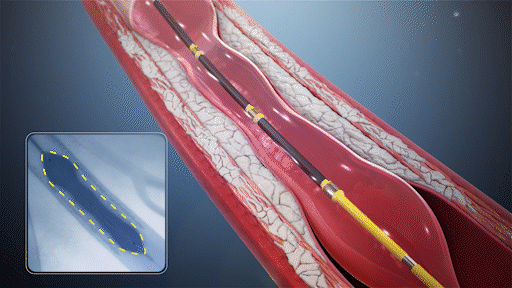
The left anterior descending artery was the toughest lesion. The balloon was inserted into the lesion without difficulty. After filling the balloon, this clearly showed the classic dog bone effect, or bowtie appearance, because the balloon did not open like a straight sausage. The blockage in the middle portion did not allow the artery to expand fully. We could not eradicate the stenosis even with 20 atm of pressure and large balloons because the balloon would not fully expand. Therefore, a stent could not be placed in this lesion. The patient was placed on medical management and brought back to the hospital in August 2021 for a new treatment called intravascular lithotripsy or IVL therapy.

Click here to sign up for Dr. J's Newsletter!

This is an angiographic image of a stent in the circumflex which is the stent that is higher up in the picture, and the stenosis in the left anterior descending artery is clearly visible as a tight narrowing. Also visible, is extensive calcification of the left anterior descending artery.

Shockwave Animation

Animation courtesy of Shockwave Medical Inc.

Demonstration of Shockwave

This is a video of the catheter outside the body and the balloon is dilated first and filled up with contrast material and then the pressure waves are seen as pulses and light. My finger is not hurt because the IVL therapy does not affect normal tissue. It only affects calcification.

This is an angiogram of the contrast filling the left anterior descending artery and the stent is clearly seen, and the vessel now no longer has that tight area of stenosis

This new therapy is conducted by placing a special catheter inside a blood vessel that is calcified. Sonic pressure waves are delivered in the balloon segment of the catheter. This exposes the surrounding artery walls to the waves and pulsations, shattering the calcium in the plaques, thereby improving the blood vessel’s compliance. It is the severe calcification that prevents the artery from fully expanding. The calcium makes the blood vessel like a lead pipe. The danger of this type of situation is that excessive force applied or excessive expansion of that blood vessel can result in a dissection (tearing of the artery). Now, by inserting this special balloon and delivering this IVL therapy into the segment with the calcification and stenosis, the characteristics of the wall change. The disruption of the calcium allows expansion of the blood vessel. The initial images clearly demonstrate that the balloon had a dog bone effect. However, once IVL is used, the balloon fully expands. Following the expansion, the shockwave catheter is removed, and now the stent can be placed. The stent expansion is now possible because the balloon will dilate fully, allowing the stent to expand fully. This gives the artery a much larger lumen (the part of the blood vessel where the blood flows). The angiogram clearly demonstrates the eradication of the stenosis or blockage. This was only possible because of the shockwave therapy in a patient who had proven failure of high-pressure balloon dilations.

Another analogy often used to describe the effects of shockwave therapy is that of an ice bag. If the ice bag has been frozen for a while and is dropped onto the floor, the individual blocks of ice separate from each other and can now move. Similarly, the calcium in the blood vessel walls is shattered and separated from each other so that the smaller pieces of calcium can be expanded and pushed tightly against the wall. In the cath lab the term we use is we changed the compliance of the blood vessel from noncompliant to more compliant. It is the same as saying that the vessel could not be stretched now can be stretched.

This procedure represents a significant advancement in our ability to treat severely calcified stenosis percutaneously.

Animation courtesy of Shockwave Medical Inc.
Learn more about the Shockwave IVL System