What is Intravascular Lithotripsy?
Intravascular Lithotripsy (IVL) is a novel application of lithotripsy, an approach that has been used for decades to safely break up kidney stones. It’s now available to treat problematic calcium in the coronary arteries that can reduce blood flow in the heart.
How Does the Shockwave's IVL Technology Work?

Image and animation courtesy of Shockwave Medical Inc.
A blocked artery can occur anywhere in the body. Blockages are due to the formation of fatty plaques known as atherosclerosis. As plaque builds up in the arteries, it becomes more difficult for blood to reach the body’s tissues. When the tissue is deprived of the oxygen rick blood it needs, symptoms may occur. When this plaque ruptures, a clot will form leading to further obstruction of the artery.
Most blockages are treated with balloon angioplasty and stenting. Angioplasty is the stretching of an artery to widen it, followed by stent placement. This is known as percutaneous transluminal angioplasty (PTCA).
Since the restenosis (re-narrowing) rate is high in angioplasty, a stent is often placed to ensure blood flow through the artery.
This new LVL Therapy allows physicians to fracture the problematic calcium – using sonic pressure waves – so that the artery can be safely expanded, and blood flow is restored with the placement of a stent and without unnecessary complications. [i]
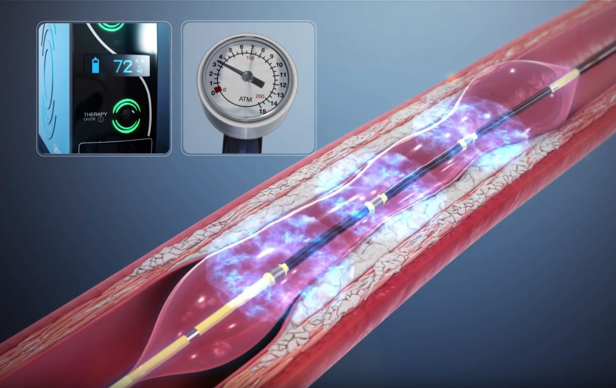
The therapy is administered by using the standard technique of delivering the catheter and inflating a balloon to a low pressure. The operator then activates the sonic pressure waves using lithotripsy to crack calcium in the artery wall. The balloon can then be expanded to prepare the lesion for the delivery of a stent.
A special catheter is placed inside a blood vessel that is calcified. Sonic pressure waves are delivered in the balloon segment of the catheter. This exposes the surrounding artery walls to the waves and pulsations, shattering the calcium in the plaques, thereby improving the blood vessel’s compliance. It is the severe calcification that prevents the artery from fully expanding. The calcium makes the blood vessel like a lead pipe. The danger of this type of situation is that excessive force applied or excessive expansion of that blood vessel can result in a dissection (tearing of the artery). Now, by inserting this special balloon and delivering this IVL therapy into the segment with the calcification and stenosis, the characteristics of the wall change. The disruption of the calcium allows expansion of the blood vessel. The initial images clearly demonstrate that the balloon had a dog bone effect. However, once IVL is used, the balloon fully expands. Following the expansion, the shockwave catheter is removed, and now the stent can be placed. The stent expansion is now possible because the balloon will dilate fully, allowing the stent to expand fully. This gives the artery a much larger lumen (the part of the blood vessel where the blood flows). The angiogram clearly demonstrates the eradication of the stenosis or blockage. This was only possible because of the shockwave therapy in a patient who had proven failure of high-pressure balloon dilations.
[i] Hill J., Kereiakes D., et al. IVL for Severely Calcified Coronary Artery Disease. J Am Coll Cardiol. 2020 Dec, 76 (22) 2635–2646. https://www.jacc.org/doi/full/10.1016/j.jacc.2020.09.603