What is an Echocardiogram?
Echocardiogram
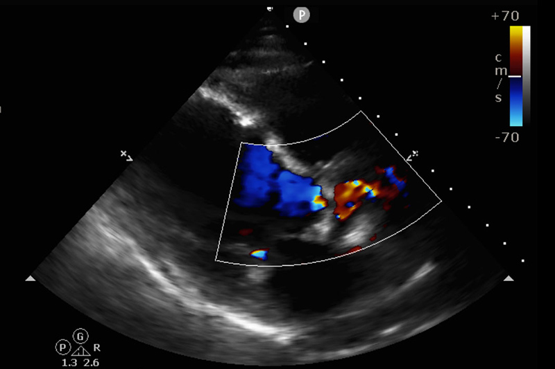
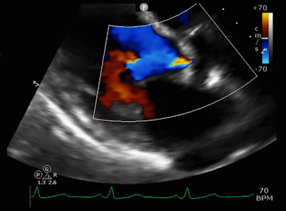
An echocardiogram (ECHO) is a simple diagnostic study used to assess the condition and function of the heart using high frequency waves.
This study is performed here in our office.
What does an Echocardiogram show?
- Congenital heart defects
- Previous heart attacks
- Blood clots in the heart
- Infection of the heart valves
- May explain chest discomfort or shortness of breath
- May explain the cause on an irregular heartbeat or heart sound
- Determines the overall strength of contractions of the heart
- Pericardial effusion (fluid around the heart)
What is the risk?
There is no risk associated with this study.
No radiation exposure is used during this study.
How do I prepare for the study?
No specific preparation is required. You may follow your daily eating habits before and after the study.
Clothing and jewelry need to be removed from the waist up to perform the study.
What happens during the study?
You will lie on a table while a trained ultrasound technician applies a clear jelly over the desired area. A probe is used to glide over this jelly which helps in transmission of sound waves through the skin.
The probe is pressed firmly over the chest to view the heart. Sound waves produced by the heart, create an echo that can be formed into an image. These images are recorded on to a monitor to be interpreted.
You may be asked to momentarily hold your breath or turn into a different position to enhance the images of the heart.
Sound waves given off the blood help to assess the direction of blood flow and blood pressures in the arteries of the heart. This also detects any abnormal sounds that may be produced by the heart.
The study may last up to 30 minutes.
What happens after the study?
After the study is over, the jelly is wiped off and you may return to your daily activities.
The result:
- The size of the heart and the chambers of the heart
- The thickness and motion of the ventricular walls
- The movement and function of the of the heart valves
- The cardiac output (volume of blood pumped out of the heart per minute)
- The pressures within various chambers of the heart and major blood vessels in the heart
- The blood flow within the heart
- The heart function changes over time
- The presence of blood clots inside the heart