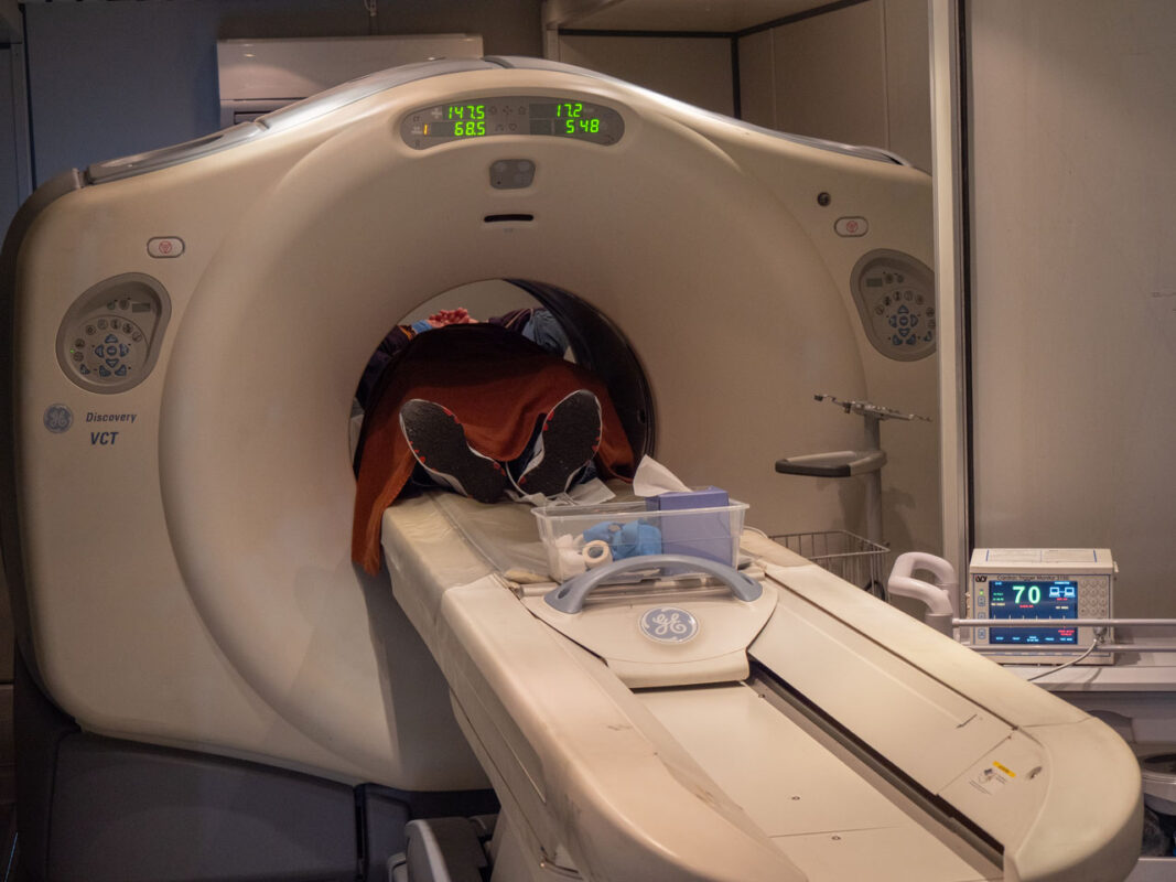
What is a Nuclear PET Scan?
A nuclear rubidium PET scan is a non-invasive nuclear imaging test used to diagnose coronary artery disease. It uses a radioactive tracer, specifically Rubidium, to produce high-resolution heart images. The tracer is injected into the bloodstream and taken up by the heart, and a PET camera detects the radiation to produce images of the heart.
Why Do I Need a Nuclear PET Scan?
A Nuclear PET Scan is performed for the following reasons:
- To evaluate the cause of chest pain and to rule out coronary artery disease.
- To determine the ability of the heart to tolerate exercise in people who have had a heart attack or heart surgery. In these patients, the PET Scan can identify new blockages as well.
- To evaluate the effectiveness of the current medical program.
- To assess blockages after coronary intervention or coronary artery bypass surgery.
- To screen for coronary artery disease in a high-risk person, such as those with high blood pressure, cholesterol, diabetes, smoking, and a family history of heart disease.
- To clarify and confirm diagnosis of coronary artery disease in situations of equivocal or borderline stress test results.
- Nuclear PET scan is the most accurate nuclear way to evaluate coronary artery disease in obese patients.
Test Preparation
- Do not eat 6 hours prior to the scan. (if patients are diabetic do not eat 4 hours before the scan.)
- No caffeine, alcohol and nicotine products 12 hours before scan.
- All medications should be taken as prescribed.
- Stay well hydrated the day prior and the day of the test, plain drinking water only the day of the test.
- Eat a high-protein, low-carbohydrate diet 24 hours before the scan.
- You may bring a light snack with you for after completion of the test.
- Wear a button-down short-sleeve shirt (no metal buttons or zippers) and athletic shoes. Do not wear necklaces. Women should NOT wear a dress.

